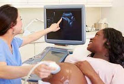
Ultrasound in Pregnancy
Ultrasound: high-frequency sound waves that travel at 10-20 million cycles per second. The pattern of echo waves results in a picture of tissue and bone. Ultrasound (also called sonogram) is a prenatal test offered to most pregnant women. It uses sound waves to show a picture of your baby in the uterus (womb). Ultrasound helps your health care provider check on your baby’s health and development.
An ultrasound is generally performed for all pregnant women around 20 weeks into their pregnancy. During this ultrasound, the doctor will confirm that the placenta is healthy and that your baby is growing properly in the uterus. The baby’s heartbeat and movement of its body, arms and legs can also be seen on the ultrasound.
If you wish to know the gender of your baby, it can usually be determined by 20 weeks. Be sure to tell the health care provider performing the ultrasound whether or not you want to know the gender of your baby. Please note that ultrasound is not a foolproof method to determine your baby’s gender; there is a chance that the ultrasound images can be misinterpreted.
In 1987, UK radiologist H.D. Meire, who had been performing pregnancy scans for 20 years, commented, “The casual observer might be forgiven for wondering why the sonography medical profession is now involved in the wholesale study of pregnant patients with machines emanating vastly different powers of one’s which is not shown to be harmless to obtain information which is not shown to be of any clinical value by operators who’re not certified as competent to perform the operations”.
Ultrasound Routine prenatal ultrasound (RPU) actually detects only between 17 and 85 percent of the 1 in 50 babies who have major abnormalities at birth. RPU can identify a low-lying placenta (placenta previa). However, 19 of 20 women who have placenta previa detected on an early scan will be needlessly worried: the placenta will effectively progress without causing problems in the birth. Furthermore, detection of placenta previa by RPU has not been found to be safer than detection in labor.
The American sonography College of Obstetricians has figured “in a population of ladies with low-risk pregnancies, neither a reduction in perinatal morbidity and mortality nor less rate of unnecessary interventions can be expected from routine diagnostic ultrasound. Thus ultrasound should be performed for specific indications in low-risk pregnancy.
Ultrasound in Pregnancy Results of ultrasound include cavitation, a process wherein the small pockets of gas that exist within mammalian tissue vibrate after which collapse. Within this situation “…temperatures of numerous thousands of degrees Celsius in the gas create a number of chemicals, some of which are potentially toxic. These violent processes might be produced by microsecond pulses from the kind that are utilized in medical diagnosis.” (American Institute of Ultrasound Medicine Bioeffects Report 1988). The value of cavitation in human tissue is unknown.
Ultrasound in Pregnancy Research has suggested these effects are of real concern in living tissues:
- Cell abnormalities caused by contact with ultrasound were seen to persist for several generations.
- In newborn rats (similar stage of development as human fetuses at 4 to 5 months in utero), ultrasound can harm the myelin that covers nerves.
- Exposing mice to dosages typical of obstetric ultrasound cased a 22% reduction in the speed of cell division and doubling of the rate of aptosis (programmed cell death), in the cells from the small intestine.
- Ultrasound in Pregnancy Two long-term randomized controlled trials comparing exposed and unexposed children’s development at eight to nine years of age found no measurable effect from ultrasound. However, the authors comment that intensities used today are lots of times greater than there have been in 1979 and 1981.
Be sure to visit the Painfree Childbirth Program Page, and know more about this popular program for eliminating fear and discomfort of labor pain from your childbirth.


